

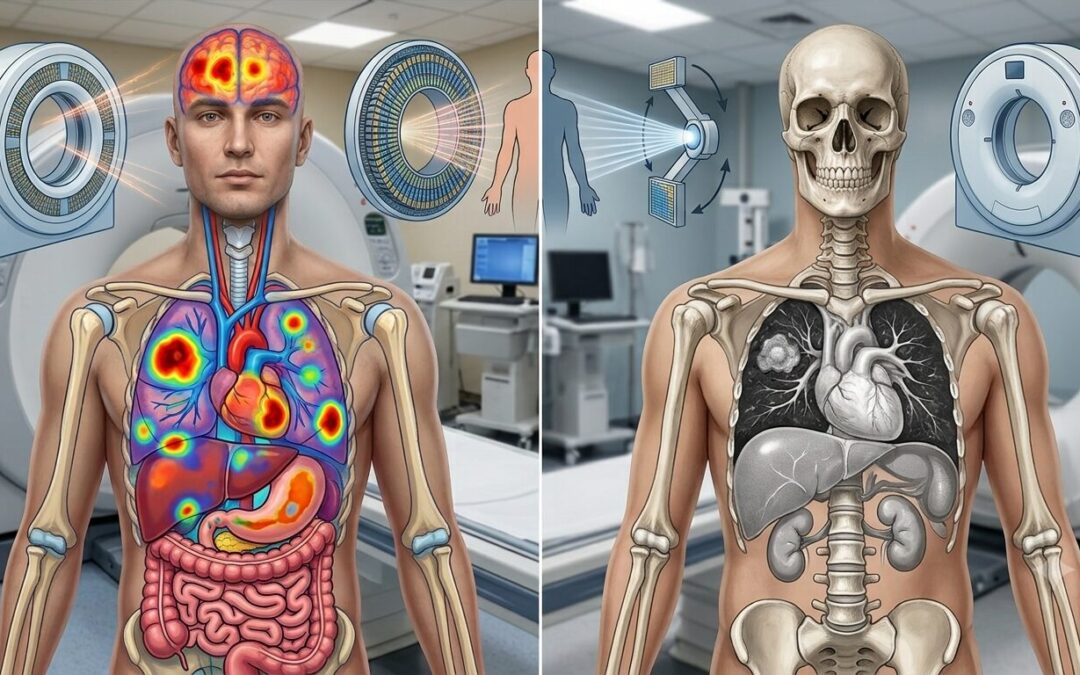
Neither is universally better, and that’s not a diplomatic non-answer. It’s the clinical reality. A CT scan shows anatomy. It maps the body’s physical structure, the size, shape, and location of organs and tumours. A PET scan shows metabolism. It reveals which cells in the body are consuming glucose at an abnormally high rate, which cancer cells typically do. One tells you where something is. The other tells you whether it’s biologically active. Used together in a combined PET/CT scan, they give a picture that neither can produce alone, structure and function in the same image, which is why PET/CT has become the standard for cancer staging and treatment monitoring in most centres.
Dr. Sandeep Nayak, Best Cancer Treatment in Bangalore, explains what each brings: “CT gives us anatomy, where the tumour is, how big it is, what it’s pressing against. PET tells us about activity. Is this lymph node actually involved or just enlarged from an old infection? Is this area lighting up because there’s active cancer or scar tissue from previous treatment? The combination is what makes the difference in clinical decision-making, particularly for staging and for assessing response after treatment.”
Trying to understand which scan your oncologist ordered and why?
PET Scan vs CT Scan: What's the Difference?
|
CT Scan |
PET Scan |
PET/CT Combined |
|
|
What It Shows |
Physical anatomy and structure |
Metabolic activity of cells |
Both structure and activity together |
|
How It Works |
X-ray beams create cross-sectional images |
Radioactive glucose tracer taken up by active cells |
CT and PET data merged into one image |
|
Best For |
Tumour size, location, organ involvement |
Active disease, lymph node involvement, spread |
Staging, restaging, treatment response |
|
Cancer Detection |
Good for structural changes |
Better for early metabolic changes |
Most accurate combination |
|
Radiation |
Moderate |
Low to moderate |
Slightly higher but combined in one session |
|
Time |
15 to 30 minutes |
2 to 3 hours including tracer uptake |
2 to 3 hours including tracer uptake |
|
Cost |
Lower |
Higher |
Higher but replaces need for both separately |
|
Widely Available |
Yes |
Less widely available |
Available at major cancer centres |
The table captures the technical differences, but in practice most oncologists order a PET/CT when staging or reassessing cancer rather than choosing between the two separately. More on how imaging feeds into treatment planning at MACS Clinic is on the precision oncology page.
When Is Each Scan Used?
The scan ordered depends on what clinical question needs answering at that point in the patient’s journey.
CT Scan Alone: Still used widely for initial assessment when a lump or symptom is being investigated. It’s faster, more accessible, and less expensive. For monitoring certain cancers where metabolic activity isn’t the primary question, tracking tumour size during chemotherapy for example, CT remains practical and sufficient. It’s also the scan of choice for guided biopsies, where the radiologist needs real-time anatomical imaging to direct a needle accurately.
PET Scan Alone: Rarely ordered without CT these days. The metabolic information without anatomical context is harder to interpret. A hotspot on a PET scan needs the CT image to tell you exactly where in the body it’s sitting and what structure it corresponds to.
PET/CT Combined: This is the standard for initial staging of most solid tumours, for restaging after treatment, and for assessing whether a treatment is working. When a patient with lung cancer needs to know whether mediastinal lymph nodes are involved, a PET/CT answers that question more reliably than CT alone. When someone who has finished chemotherapy needs to know whether residual masses are active disease or dead tissue, PET/CT is what distinguishes the two. Our blog on what is metastatic cancer covers how staging scans connect to decisions about spread and treatment.
MRI as an Alternative: Worth mentioning because it comes up often. MRI doesn’t use radiation and gives superior soft tissue detail in certain areas, particularly the brain, liver, and pelvis. For rectal cancer staging, brain metastasis assessment, and certain liver lesions, MRI is preferred over CT or PET/CT specifically because of its soft tissue resolution.
After Treatment Monitoring: Liquid biopsy and ctDNA testing are increasingly used alongside imaging to monitor treatment response, catching molecular signs of disease activity before they show up on any scan. Imaging and molecular monitoring together give a more complete picture than either alone.
Why Choose MACS Clinic for Cancer Diagnosis and Staging?
Dr. Sandeep Nayak’s team at MACS Clinic treats imaging as one part of a complete diagnostic picture, not the whole story. PET/CT findings are reviewed alongside biopsy results, molecular profiling, and clinical examination before any staging conclusion is reached or treatment plan finalised. A hotspot on a PET scan doesn’t automatically mean active cancer. A normal CT doesn’t rule out microscopic disease. Getting the interpretation right matters as much as getting the scan done.
For patients who’ve had imaging elsewhere and want a second read in the context of a full oncology assessment, the team reviews all available scans as part of the initial consultation. Those who want to discuss their imaging results or understand what comes next can reach the team at +91 9482202240.
FAQs
Is a PET scan always better than a CT scan for cancer?
Depends on what the doctor needs to know. CT handles structure, size, and guided biopsies well. PET/CT wins for staging and checking whether treatment is working. Neither replaces the other.
Can a PET scan miss cancer??
Yes. Slow-growing or well-differentiated tumours don’t always consume enough glucose to show up. A clear PET scan is reassuring but not a guarantee.
How long does a PET/CT scan take?
Around two to three hours in total. Most of that is waiting for the tracer to absorb. The actual scan is only 20 to 30 minutes.
Is the radiation from a PET/CT safe?
Higher than a CT alone, yes. But for someone being staged or monitored for cancer, the clinical value far outweighs the exposure. It’s not ordered without a good reason.
Disclaimer: This content is published for educational and informational purposes only.