
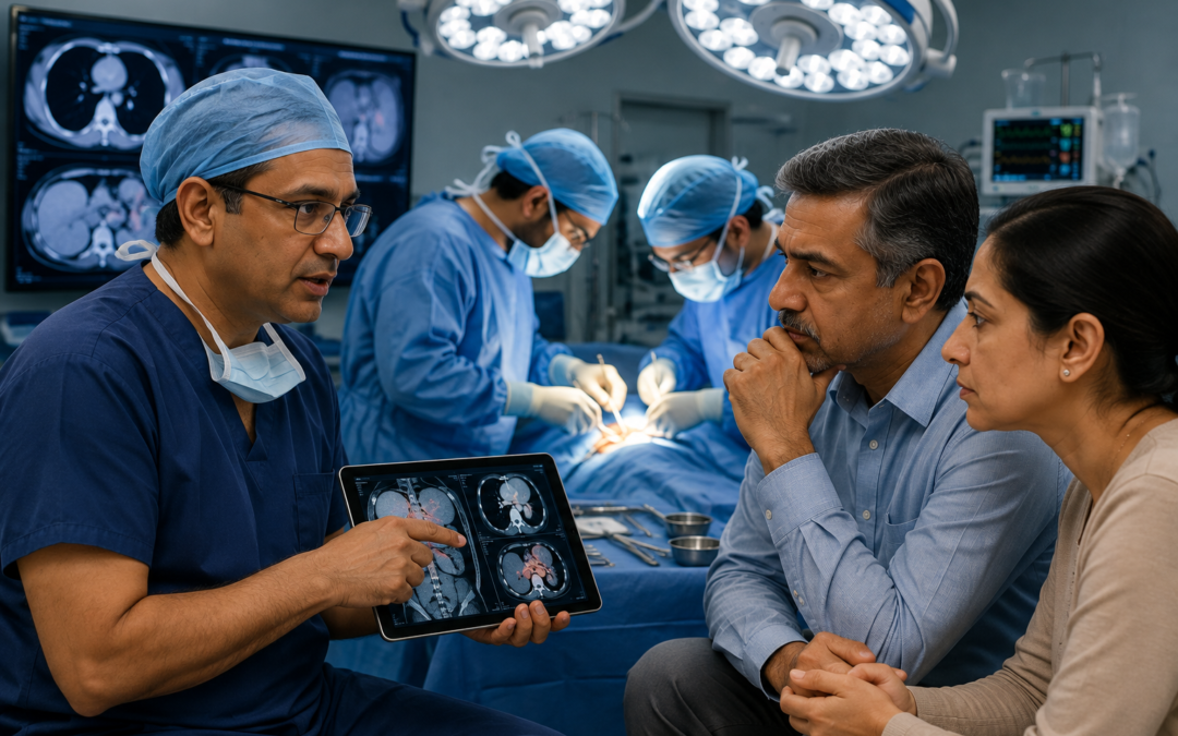
Scans are good. They’re not perfect. A CT, MRI, or PET scan tells the surgical team what it can detect at the resolution it operates at and cancer doesn’t always cooperate with that resolution. Microscopic spread along a surface, small deposits tucked behind a structure, lymph nodes that look borderline on imaging but are clearly involved when held in a surgeon’s hand none of these reliably show up before the operation. So sometimes, the surgeon opens up and finds more than anyone expected.It’s not a failure of the pre-operative workup. It’s the reality of what imaging can and cannot do. What matters is what happens next and that depends on what was found, where it was, and what the surgical team decides at the moment.
According to Dr. Sandeep Nayak, Best Cancer Treatment in Bangalore, “Finding more disease at surgery than the scans suggested happens more than patients realise. The question isn’t whether it happens — it’s whether the team in that operating room is equipped to make good decisions about it when it does. Sometimes we remove more. Sometimes we stop, close, and reassess with the full pathology picture before committing to a bigger operation. The decision is made based on what we find, not what we planned.”
Discovered unexpected cancer spread during surgery?
What the Surgeon Actually Does When More Cancer Is Found?
The intraoperative decision depends on what’s found and whether removing it safely is possible.
Remove as much as safely possible
When the additional disease is localised a few extra deposits, a lymph node group more involved than imaging suggested the surgeon typically extends the resection to include it, provided that doesn’t put critical structures at risk. More complete removal generally means better outcomes, and the operating room is the best opportunity to achieve that.
Stop and reassess when the extent is beyond what’s safe
When what’s found is significantly more than expected widespread peritoneal deposits, involvement of major vessels, disease in areas that weren’t planned for the safer decision is sometimes to stop the primary resection, take biopsies to confirm what’s present, and close. Forcing a larger operation than planned in a single sitting, without the team prepared for it, risks serious complications for uncertain benefit. The patient then gets restaged based on the intraoperative findings and the full pathology, and a revised plan is built from there.
Use frozen section pathology intraoperatively
Most experienced surgical oncology centres have access to frozen section analysis during the operation a rapid pathology assessment of tissue taken from suspicious areas that returns results within minutes. This allows the team to confirm whether what they’re looking at is genuinely malignant before deciding whether to extend the operation or stop. It’s one of the most important tools available when the intraoperative picture diverges from what the scans showed.
Update staging and revise the treatment plan after surgery
Whatever is found, the post-operative plan changes to reflect it. A patient who goes in staged at Stage 2 and comes out with intraoperative findings consistent with Stage 3 or 4 disease gets restaged formally, and the cancer treatment plan adjuvant chemotherapy, radiation, targeted therapy — is revised accordingly. Surgery is one data point in a longer treatment course, not the whole story.
Why Scans Don't Always Show the Full Picture?
Understanding why this happens is as important as understanding what to do about it.
Imaging has resolution limits. A CT scan picks up lesions reliably above a certain size roughly 5 to 8mm depending on location. Below that threshold, disease can be present and invisible. Peritoneal deposits in particular are notoriously difficult to characterise on cross-sectional imaging, which is why staging laparoscopy before major peritoneal surgery exists as a separate step in some cancers.
Some structures are difficult to assess radiologically. Lymph nodes are evaluated on size criteria on CT enlarged means suspicious, normal-sized means likely clear. But normal-sized nodes can carry microscopic metastatic disease, and enlarged nodes can be reactive rather than malignant. The only reliable way to know is pathology, which requires tissue.
Tumour biology can change between imaging and surgery. A PET-CT done six weeks before the operation is six weeks old by the time the patient is on the table. Fast-growing tumours can progress meaningfully in that window. For a detailed look at how staging is done before surgery and what each modality is actually trying to detect, the previous blog on How Cancer Staging Is Done Before Surgery covers the full workup process.
Why Choose MACS Clinic for Cancer Surgery?
Dr. Sandeep Nayak’s team at MACS Clinic plans every cancer operation through tumour board review before the date is confirmed — which means the intraoperative decision-making framework is established before the patient is on the table, not improvised in the moment. Frozen section pathology is used where it changes surgical decision-making. Where staging laparoscopy reduces the risk of an unnecessary major operation, that step is done first.
Finding more disease than expected is a clinical reality. What separates good outcomes from poor ones is the team’s ability to make sound decisions about it when it happens. Those who want to discuss their case can reach the team at +91 8035740000.
FAQs
Does finding more cancer during surgery mean the original plan failed?
Not at all. Imaging has resolution limits that surgery doesn’t. Finding more disease intraoperatively is a known and expected possibility in cancer surgery, not a sign that the pre-operative workup was inadequate.
Will the surgery be extended to remove the extra cancer found?
Sometimes yes, sometimes no. It depends on where the additional disease is and whether removing it safely is feasible in that sitting. The surgeon makes that call based on what’s present and what the risk of extending the operation would be.
What happens to the treatment plan if more cancer is found?
It gets revised. Post-operative staging is updated based on intraoperative and pathology findings, and the plan for chemotherapy, radiation, or targeted therapy is adjusted to reflect what was actually found rather than what imaging predicted.
Should I ask my surgeon about this possibility before the operation?
Yes. Asking what happens if more disease is found than expected is a completely reasonable pre-operative question. The answer tells you how prepared the team is for that scenario.
Disclaimer: This content is published for educational and informational purposes only.