Breast Cancer Treatment in Bangalore

Breast cancer is one of the most common types of cancer in women. Every year, approximately 1,000,00 women in India are diagnosed with breast cancer. Although the disease is widespread, many women are unaware of it until it is too late.

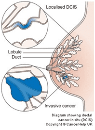
Breast cancer develops in the cells of the breast. It usually starts in the breast lobules or ducts.

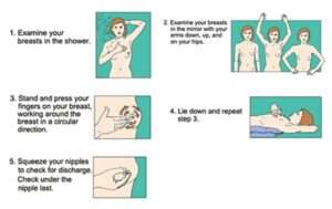
Because breast cancer often shows symptoms, it is critical to self-examine your body and observe any unusual indicators. Something other than cancer could be causing your symptoms. Hence, the only way to be sure is to visit your doctor.

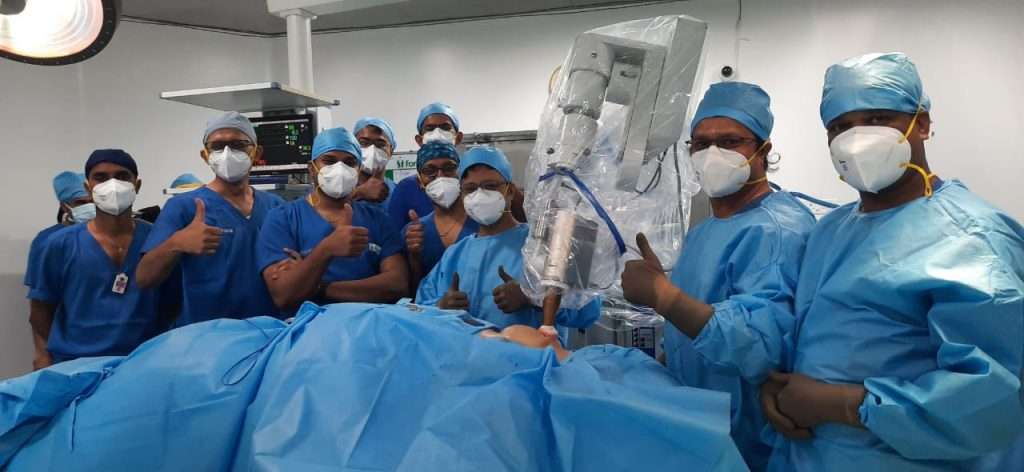
At MACS Clinic, Dr. Sandeep Nayak, with his well-trained team, offers advanced and customized breast cancer treatment in Bangalore, India.

MACS Clinic is regarded as one of the best cancer hospital in Bangalore because we are committed to advancing the diagnosis and treatment of a wide range of malignancies, including breast cancer.

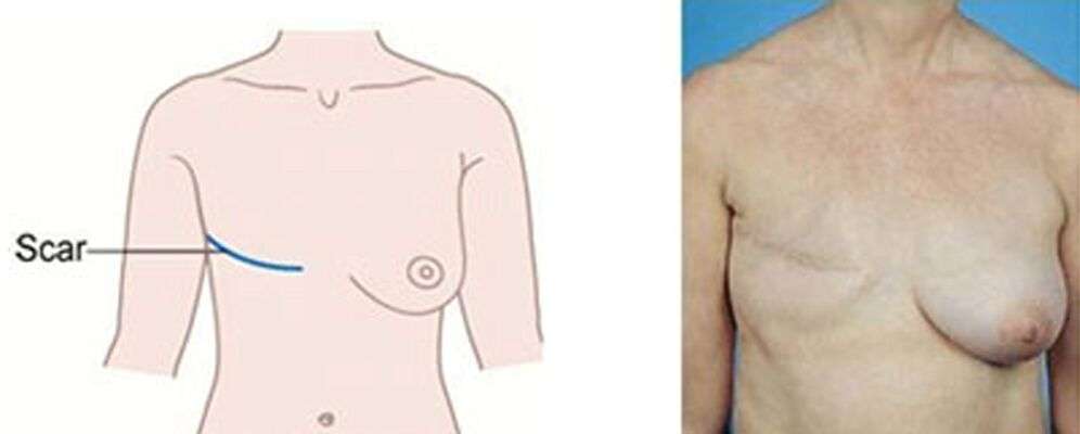
Our clinic is equipped with minimally-invasive surgical techniques and single-day breast cancer radiation therapies like IORT. These methods help the patients recover quickly and offer long-term results.

This article can help if you want to learn more about breast cancer symptoms, diagnosis, and treatments. Continue scrolling to know more.

Now, let’s talk about Breast Cancer Symptoms

Breast cancer signs and symptoms differ from one woman to the next. However, most people may experience mild pain or a lump in their breasts.
In its early stages, breast cancer may not show any signs. A tumour is too small to feel in many cases, but an abnormality on a mammogram can be seen.

Breast cancer symptoms include:

  • mass or lump
  • puckering
  • soreness
  • dimpling
  • nipple bulging
  • change in skin complexion

The presence of one or more of these signs does not imply that you have breast cancer. Thus, you should seek the advice of a specialist.

Diagnosis of Breast Cancer

Your doctor will first perform a physical examination. It entails inspecting your breasts for lumps, discharge from one or both nipples, changes in size or shape of the breast, nipple changes such as puckering, and inflammation of the skin surrounding the nipple.
In addition, the doctor may inquire as to whether you are experiencing pain in one area of your chest wall and/or if there have been any changes in your periods.

Other diagnostic tests may include:

Mammogram:
It detects changes in breast tissue using low-energy X-rays. This test can aid in the detection of potential tumours before they become lethal. A doctor examines the mammogram and reports any abnormalities.

Breast Cancer Treatment:
Surgery, radiation therapy, chemotherapy, or a combination of these are all options for breast cancer treatment. However, surgery is a must for curing breast cancer. The type of surgery selected depends on the stage of the cancer.

Breast ultrasound:
Using sound waves, a breast ultrasound creates a picture of the tissues deep within your breast. It can help your doctor distinguish between a solid mass, such as a tumour, and a benign cyst.

If your doctor suspects you have cancer, they may suggest that you have tests like an MRI scan or a breast biopsy to confirm the diagnosis. A needle test (FNAC) or needle biopsy can provide a diagnosis in most cases.

Conventional Breast Cancer Surgery

Radical mastectomy and modified radical mastectomy are the two conventional breast cancer surgeries.

Radical mastectomy

It involves the removal of the breast, chest muscles, and all of the lymph nodes under the armpit. Though this was a standard procedure 80 years ago, presently, it is used only when the tumor has spread to the chest muscles. This is a rare occurrence nowadays, as patients tend to come early.

This surgery has many side effects, and research has proven that this breast cancer surgery does not benefit the patient. So, this procedure is rarely required.

Modified radical mastectomy (MRM)

It has been the standard of care for breast cancer for many years. This procedure is less traumatic than radical mastectomy. It entails removing the breast, affected lymph nodes under the arm, and the lining over the chest muscles. This reduces the discomfort to the patient while providing an adequate cure.

This surgery is performed only when it is impossible to save the breast-by-breast conservation therapy or lumpectomy. However, in those patients who need a mastectomy, the breast can be reconstructed in the same sitting or after completing all treatments.

Advanced Breast Cancer Surgery

Minimally Invasive Breast Surgery (MIBS):

Over the past 25-30 years, the trend toward saving normal, the natural breast has increased, supported by robust research. It has been proven that preserving breasts DOES NOT affect the cure rate (oncological outcome).

Today increasing numbers of patients can undergo one of the types of minimally invasive breast cancer surgery and save their natural breast, improving the cosmetic outcome and quality of life.

Skin Sparing Mastectomy and Reconstruction:

It is the most advanced form of breast surgery performed very selectively at only a few centers. This is mainly performed for patients who are eligible for nipple-areola skin-sparing mastectomy (extensive changes on mammography, multicentric cancer, or prophylactic mastectomy for high genetic risks).

The entire surgery, including flap reconstruction, can be performed with very tiny wounds with the least possible pain and scaring. The role of robotic surgery is predominantly in reconstruction in these cases.

Some cases require the removal of entire breast tissue along with the nipple. This happens in large breast cancers or multiple cancer cases in the same breast. The skin over the breast is softer, and replaced skin from other body parts cannot match that skin.

Retaining natural skin over the breast gives the best cosmetic results compared to any other form of reconstruction (tissue transfers). The breast can be reconstructed entirely through the same incision or wound to restore the normal shape and contour in the same sitting.

The nipple can be reconstructed using tattooing. A few specific flaps are commonly used for the reconstruction of the breast. These are TRAM flap, LD flap, or DIEP flap. The use depends on various situations.

Breast Conservation Surgery or Lumpectomy with Oncoplastic Surgery:

It involves the removal of the cancer lump or tissue in the breast and some of the normal breast tissue around it and the lining over the chest muscles below, clearing all the cancer-bearing tissue. Usually, some lymph nodes under the arm are taken out (through a separate incision) and tested for the possible spread of cancer.

The oncoplastic surgery technique helps give the normal breast contour (shape) after adequate removal of cancer tissue. The surgical options may include:

  • Local tissue rotation (rotation mammoplasty).
  • Tissue transfer from a distant body area (Latissimus Myocutaneous Flap).
  • A silicone prosthesis.

The patient gets almost a normal breast appearance at the end of this.

Nipple Areola Skin Sparing Mastectomy and Reconstruction :

Some cases require the removal of entire breast tissue without the need to remove the nipple (extensive changes on mammography, multicentric cancer, or prophylactic mastectomy for high genetic risks like BRCA mutation families). Skin-sparing and nipple-sparing mastectomy is routinely performed through one or two short incisions placed over the breast.

The breast can be reconstructed entirely through the same wound to restore the normal shape and contour in the same sitting using one of the tissue flaps (from the back or belly). This gives almost normal looks to the breast.

Robotic Breast Surgery:

It is the most advanced form of breast surgery performed very selectively at only a few centers. This is mainly performed for patients who are eligible for nipple-areola skin-sparing mastectomy (extensive changes on mammography, multicentric cancer, or prophylactic mastectomy for high genetic risks).

The entire surgery, including flap reconstruction, can be performed with very tiny wounds with the least possible pain and scaring. The role of robotic surgery is predominantly in reconstruction in these cases.

Armpit (Axillary) Lymph Nodes

Sentinel Node Biopsy (SNB):

Today, it is the procedure of choice for managing the armpit lymph nodes in almost all breast cancers. Only the nodes that drain the breast are selectively removed, avoiding the nodes draining the arm. This way, the risk of arm swelling in the future comes down to less than 2% from 40% when all armpit nodes are removed. This is a big difference for the patient.

We perform this procedure using the double dye technique so that nodes are not missed. We use methylene blue and indocyanine green (ICG). Some centres perform this by using a radioactive substance. However, we have been able to avoid using it effectively.

Axillary Lymph Node Dissection (ALND):

Every breast cancer surgery involves removing the lump and draining lymph nodes in the armpit or axilla. The nodes in the axilla are divided into three levels, and at least 2 of these levels are removed while performing ALND surgery.

The unfortunate part of this surgery is that about 40% of the patients develop swelling of their hands in their lifetime as the lymph nodes responsible for the arm and breast are in the axilla. It is a very high number. This was a standard of care for all breast cancers until about 15 years back. Even today, ALND is required for some of the patients who come to us with involved nodes.

Axillary Reverse Mapping (ARM):

Every patient is not fortunate enough to reach us at an early stage. There are times when patients have involved lymph nodes in the armpit. Traditionally all these patients have to undergo ALND and have a very high lifetime risk of arm swelling up.

We have devised an axillary reverse mapping technique to identify the lymphatic ducts coming from the arm and preserve those, removing all the lymph nodes. By doing this, we may be able to reduce the risk of arm oedema or swelling.

IORT: A Way to Complete Breast Cancer Treatment in One Single Day

Breast cancer is one of the most common cancers in the world. However, it is also one of the most researched cancers globally, and things have changed enormously over the years. Today in most patients, we can cure cancer, save the breast, and even complete the treatment in one day by doing IORT. We are fortunate to be living in this age where there are many possibilities, and the cure rate is very high.

One thing that deters patients from undergoing cancer treatment for breast cancer is that they need to go for treatment for a prolonged duration of time. However, newer technology has changed it for some early breast cancers.
According to a study published in the British Medical Journal, a single dose of radiation therapy known as intraoperative radiation therapy (IORT) administered during a lumpectomy produced roughly comparable results for early-stage breast cancer as traditional whole-breast radiation therapy administered after surgery. So, 30 days of treatment can be completed in 30 minutes.

Traditional Whole-Breast Radiation Therapy

Traditionally, whole-breast radiation therapy has been used to remove breast cancer in most patients with a lumpectomy or breast conservation surgery. Radiation after a lumpectomy helps destroy any cancer cells that may remain in the breast after surgery.
As the name implies, whole-breast radiation therapy aims to direct a radiation beam at the entire breast suffering from cancer. Studies have shown traditional radiation to effectively reduce cancer recurrence in both the short and long term.

The disadvantages of conventional radiation therapy include regular visits to the hospital, often five days a week for three to six weeks. Maintaining this schedule can be challenging for many people, as even the caregiver’s time is required. Traditional radiation therapy may also expose healthy tissue to more radiation, such as the heart and lungs.

The goal of IORT is to avoid these issues while producing the same results.

Now, let’s know more about,

What Exactly is Intraoperative Radiation Therapy (IORT)?

Most breast cancers come back in the breast tissue very close to first cancer. IORT is given during lumpectomy surgery for 20-40 minutes, right after the cancer is removed while the patient is still under anaesthesia. While the underlying breast tissue is still exposed, a single, high dose of radiation is given directly to the area where the cancer was.

In addition to being more convenient than traditional radiation therapy schedules, the researchers who developed IORT believe it can avoid exposing healthy tissue to radiation. More than 250 centres worldwide have adopted IORT and are showing the benefit to the patients.

Let's know the Advantages of IORT
  • Quick completion of treatment in 1 day
  • Adds only 20-40 mins. to surgery
  • High precision
  • No radiation to normal tissue
  • No pain or complications from radiation
  • No difference in cancer cure results
Who is eligible for the single-day IORT treatment?

With IORT, it is possible to give single-day therapy for breast cancer in selected cases.

Case Selection (Internationally Accepted Guideline):
  • Women > 50 years old
  • Tumours measuring 2 cm or less
  • HR+
  • Without lymphovascular invasion
  • No nodal involvement
  • Negative margins

For these patients, we can perform the lumpectomy procedure along with sentinel node biopsy for lymph nodes followed by IORT. Traditionally this would take approximately a few months (about 8-12 weeks) to complete as there has to be a gap between surgery and radiation. By doing IORT (in 20-40 min), the entire process is completed in 1 day.

Can IORT be used in all breast cancer cases?

Many breast cancer patients may not be eligible for single-dose IORT due to various factors and may need the traditional whole breast radiation therapy. However, even among these patients, there is a definite scope and advantage of using IORT. There are many scenarios the benefits that have been described in these patients as well.

Along with whole breast radiation, the oncologist usually administers an additional radiation boost to the area where the cancer was there. This can be done more effectively using IORT with the following advantages:

IORT as a boost to the tumour area offers better precision

Oncoplastic reconstruction is used for cosmetic purposes. The breast tissue is moved to close the gap, or tissues from other body parts are used. In this scenario, the original breast tissue is difficult to identify. When we give IORT at the time of surgery, there is no confusion about the tumor’s location. The radiation can be delivered precisely in the presence of an operating surgeon. It eliminates the radiation error.

• Saves 4-8 days of tumor bed boost.

• Can also be considered in previously radiated patients as conventional EBRT may be difficult.

So, every breast cancer patient can benefit from opting for IORT, even when they need additional radiation.

Breast Cancer Early Detection & Prevention

Early detection is the best way to get a cure. Most early cancer patients can save their breasts. There are three steps to early detection of breast cancer.

  • Breast Self Examination: Monthly breast self-examination is one of the critical elements in detecting breast cancer early. This makes you aware of the consistency and feel of your breast. Women should perform the examination only once a month. The seventh day after your menstrual period would be the ideal day. Menopausal women should choose any one date of a month. Also, examine your armpits for any nodules. If you feel any new nodules, consult your doctor.

  • Clinical Examination: A yearly breast examination by your doctor to look for any abnormalities would help detect any lumps that you miss. These lumps that are detected need further workup to understand their nature.
  • Mammography: It is the X-ray examination of the breast. If this detects any defects, the doctor investigates the defect to see further if it is cancer. Often, oncologists suggest mammography only for women older than 40. Younger women can be examined using an ultrasound scan of the breast (sono-mammography) if there is a need to do so.
Strategies to reduce the risk of cancer:

Cancer, to a certain extent, is a lifestyle disease. Some recommendations that could help reduce the risk of cancer are as follows:

  • Develop a healthy eating habit
  • Maintain healthy weight
  • Include some physical activity in daily routine
  • Say NO to tobacco and alcohol
  • Breastfeed your baby at least for one year, as it reduces the risk of breast cancer
  • Avoid hormone replacement therapy

FAQs

What are the risk factors associated with breast cancer?

Following are some of the breast cancer risk factors:

  • Age
  • Heredity
  • Medical history of family
  • Excessive exposure to radiation or specific chemicals
  • Diet
  • Use of oestrogen-boosting hormones or medications, such as birth control pills or hormone replacement therapy pills
  • Obesity or a lack of exercise

Does wearing a padded bra increase the risk of breast cancer?

The relationship between bras and breast cancer has sparked controversy. However, no research has discovered that wearing a padded bra causes cancer.

Does treating early-stage breast cancer increase survival?

Breast cancer in its early stages can be cured with surgery, radiation, and, in some cases, chemotherapy. According to studies, breast cancer patients who receive treatment in the early stages live for five years or longer after diagnosis.

Where does breast cancer begin to spread?

Breast cancer is most likely to spread to the lymph nodes under your arm first. It could also spread to the tissue that surrounds your breast, such as your chest, collarbone, or lower neck.