

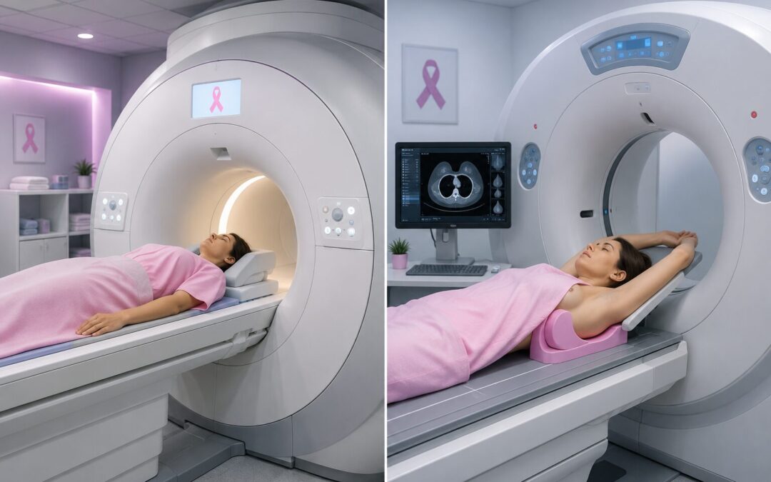
MRI is generally more sensitive, yes. But that one sentence doesn’t tell the full story and it’s not how oncologists make this decision in practice. Breast MRI picks up small tumours and early-stage cancers that ultrasound and even mammograms can miss, particularly in dense breast tissue. Ultrasound is faster, more widely available, cheaper, and handles specific jobs that MRI simply isn’t built for. Neither one replaces a mammogram as the standard screening tool. Which scan gets ordered depends on what the clinical question actually is, what the patient’s risk profile looks like, and what the breast tissue is doing.
Dr. Sandeep Nayak,who provides Best Cancer Treatment in Bangalore, breaks it down: “MRI and ultrasound are not competing tools. They answer different questions. Ultrasound tells us whether a lump is solid or fluid-filled and guides biopsy needles in real time. MRI tells us the full extent of disease in a breast, whether there are additional sites we haven’t found yet, and how the other breast looks. Ordering the right scan at the right moment is part of how diagnosis and surgical planning come together properly.”
Noticed a lump or been told your breast tissue is dense?
MRI vs Ultrasound for Breast Cancer: What's the Difference?
|
Breast MRI |
Breast Ultrasound |
|
|
Sensitivity |
Very high, 90 to 95% |
Moderate, 70 to 80% |
|
Best For |
High-risk screening, extent of disease, dense breasts |
Characterising lumps, guiding biopsy, younger women |
|
Radiation |
None |
None |
|
Contrast Agent |
Yes, gadolinium injection needed |
No |
|
Time |
45 to 60 minutes |
15 to 30 minutes |
|
Cost |
Higher |
Lower |
|
Availability |
Major centres only |
Widely available |
|
False Positives |
Higher, leads to more unnecessary biopsies |
Lower |
|
Replaces Mammogram |
No |
No |
|
Guided Biopsy |
Possible but complex |
Yes, standard use |
Higher sensitivity sounds like a clear win. It isn’t always. More sensitivity brings more false alarms, more callbacks, more biopsies on lumps that turn out to be nothing. That trade-off is real and it’s exactly why MRI isn’t the default scan for every patient who walks in. More on how imaging connects to diagnosis and surgical planning at MACS Clinic is on the breast cancer treatment page.
When Is Each Scan Actually Used?
It changes depending on where the patient is in their journey. Same woman, different question, different scan.
Ultrasound First: Something’s been felt or a mammogram flagged something. Ultrasound goes in next. It tells the radiologist within minutes whether that lump is a harmless cyst or something that needs a closer look. For women under 35 with dense tissue, it’s often the starting point because mammogram can’t read through density properly. It also guides biopsy needles in real time.
MRI When More Is Needed: Once breast cancer is confirmed, MRI maps the full picture before surgery is planned. Is it one spot or several? Does the other breast look clear? A mammogram and ultrasound together can’t always answer those questions. For women with BRCA mutations or a lifetime risk above 20%, annual MRI runs alongside mammogram because it catches what mammogram misses in dense tissue. Our blog on hereditary breast cancer covers who falls into the high-risk group where that protocol changes.
Surgical Planning: MRI findings feed directly into whether lumpectomy is realistic or whether the disease extent makes mastectomy more appropriate. Our blog on lumpectomy vs mastectomy covers how imaging shapes that decision.
Implants: MRI is the standard for checking implant integrity and for screening women where mammogram is limited by the implant itself.
Why Choose MACS Clinic for Breast Cancer Diagnosis?
Dr. Sandeep Nayak’s team at MACS Clinic picks the scan that answers the question on the table, not the fanciest one or the quickest one. Newly diagnosed patients get MRI findings reviewed alongside mammogram, ultrasound, biopsy, and receptor results before anyone talks surgery. Women in high-risk surveillance get an imaging protocol built around their personal risk profile, not a one-size schedule.
Getting the extent of disease right before the operation changes what’s possible in breast cancer surgeries. The imaging has to be right first. To discuss a lump, dense tissue finding, or a recent scan result, the team can be reached at +91 9482202240.
FAQs
Can ultrasound pick up cancer that mammogram missed?
It happens, yes. Dense breast tissue is the main reason. Mammogram has a harder time reading through it, and ultrasound fills that gap. Younger women especially tend to get both for exactly this reason.
Does a clear breast MRI mean there's no cancer?
Mostly reassuring, not completely conclusive. MRI misses things occasionally and throws up false alarms fairly often too. Women get called back for biopsies that turn out fine. A clear result is good news, not a guarantee.
Who needs annual MRI on top of mammogram?
Not everyone. BRCA carriers are the clearest group. After that, women whose overall lifetime risk sits above 20% and anyone who went through chest radiation before their thirties. If none of those apply, a yearly mammogram is still what the guidelines recommend.
Is breast MRI uncomfortable?
The scan itself isn’t painful. Lying face-down in an enclosed scanner for up to an hour is what people find difficult, especially anyone who doesn’t love tight spaces. A contrast injection goes into the arm before scanning starts.
Disclaimer: This content is published for educational and informational purposes only.