

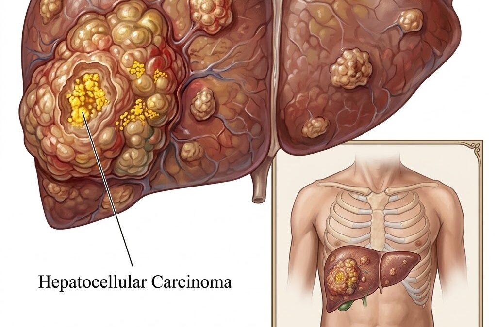
CC is the most common cancer that starts in the liver itself. Not secondary cancer that spreads there from somewhere else. This one begins in the hepatocytes and stays liver-primary. It accounts for 75% to 85% of all liver cancer cases and almost never appears in a liver that’s been healthy. Something damages it first, usually over years, often quietly. By the time symptoms appear, the window for cure has usually already closed.
According to Dr. Sandeep Nayak, Best cancer treatment in Bangalore, “Hepatocellular carcinoma is one of the few cancers where we know the risk population well in advance. Patients with cirrhosis, chronic hepatitis B or C, or NASH-related liver disease should be on a surveillance programme with ultrasound and AFP every six months. The cancers we find through surveillance are the ones we can still cure. The ones we find because of symptoms usually aren’t.”
Have chronic liver disease and not yet on a surveillance programme? That gap between who should be monitored and who actually is costs lives
What Actually Causes Hepatocellular Carcinoma?
HCC rarely appears without a reason. The liver has usually been fighting something for years before the cancer shows up.
- Hepatitis B and C:
Between them, these two viruses drive 70% to 80% of HCC cases globally. Hepatitis B is particularly dangerous because it can cause liver cancer even before cirrhosis sets in. So HBsAg-positive patients need surveillance regardless of whether their liver has scarred yet. Hepatitis C usually causes HCC through cirrhosis first, but the endpoint is the same. - Cirrhosis from Any Source:
Whether it’s alcohol, autoimmune hepatitis, primary biliary cholangitis, or something else, a cirrhotic liver is one where cells regenerate constantly and accumulate errors with each cycle. Around 80% of HCC cases develop in cirrhotic livers. The specific cause of the cirrhosis matters less than the fact of it. - Fatty Liver Disease:
MASLD, which most people still know as NAFLD, is now the fastest-growing HCC risk factor. It’s becoming more common in people who don’t drink, don’t have hepatitis, and don’t know their liver is in trouble. A lot of MASLD-related HCC is found late because nobody thought to look. - Aflatoxin Exposure:
Aspergillus moulds on stored grain and groundnuts produce aflatoxin B1, a carcinogen that causes a specific TP53 mutation and drives HCC in tropical and subtropical regions. This is one of the few pathways where liver cancer can develop without established cirrhosis, often in younger patients.
Precision Oncology molecular profiling in HCC identifies TP53, CTNNB1, and TERT mutations that guide systemic treatment decisions in unresectable cases where atezolizumab-bevacizumab is now first-line.
How Is HCC Diagnosed and Staged?
One thing that makes HCC unusual is that it often doesn’t need a biopsy to confirm the diagnosis.
- Imaging First:
In a patient with known cirrhosis, a liver lesion that enhances on arterial phase and washes out on venous phase CT or MRI is treated as HCC without tissue confirmation. That imaging pattern reflects the tumour’s blood supply being hepatic artery-dominant rather than portal vein-dominant. Standard for all lesions over 1 cm in a cirrhotic liver. - AFP Surveillance:
Alpha-fetoprotein is elevated in 60% to 70% of HCC cases. Not sensitive enough to diagnose on its own but combined with six-monthly ultrasound it catches early-stage disease in at-risk patients. A consistently rising AFP in a cirrhotic patient needs imaging immediately, not at the next scheduled appointment. - BCLC Staging:
The Barcelona Clinic Liver Cancer system stages HCC based on tumour burden, liver function by Child-Pugh score, and performance status together. It’s the staging system most treatment guidelines follow because it integrates all three factors rather than just tumour size, which alone doesn’t tell you much in a disease where the organ’s reserve is half the clinical picture. - Liver Reserve Assessment:
A patient can have a technically resectable tumour but still not be a surgical candidate if removing it would leave too little functioning liver behind. Child-Pugh score and indocyanine green retention test measure what’s left after resection. That assessment happens before surgery is offered, not after the operation reveals a problem.
Our previous blog on RABIT Thyroid Surgery is worth a read for context on how surgical planning in oncology always weighs what’s removed against what the body can sustain. In HCC, that calculation is more critical than in almost any other solid tumour.
Why Choose MACS Clinic for Hepatocellular Carcinoma?
Dr. Sandeep Nayak’s team at MACS Clinic reviews every HCC case through a multidisciplinary process that covers tumour burden, liver function, portal hypertension, and whether the patient meets criteria for resection, ablation, TACE, or systemic therapy before any recommendation is made. HCC is one of the cancers where the organ the tumour lives in shapes what’s possible at least as much as the tumour itself does.
Early-stage HCC in a cirrhotic liver needs both an oncological assessment and a hepatological one. Both need to agree on what the liver can safely tolerate before anything proceeds. Those who want to discuss their specific case can reach the team at +91 8035740000.
FAQs
What is the most common cause of hepatocellular carcinoma?
Globally, hepatitis B and C between them drive most cases. In India, hepatitis B is a particularly significant driver. Fatty liver disease is now the fastest-growing cause in people without viral hepatitis.
Can HCC develop without cirrhosis?
Yes, but it’s uncommon. Hepatitis B can cause it before significant scarring develops. Aflatoxin exposure is another route. But 80% of cases still show up in cirrhotic livers.
How is HCC diagnosed?
In cirrhotic patients, the right imaging pattern on CT or MRI is enough without a biopsy. AFP monitoring with six-monthly ultrasound is the standard surveillance protocol for at-risk patients.
Who needs liver cancer surveillance?
Anyone with cirrhosis from any cause, chronic hepatitis B carriers regardless of fibrosis stage, and patients with advanced MASLD-related liver disease. Six-monthly ultrasound with AFP is the standard.
Disclaimer: This content is published for educational and informational purposes only.